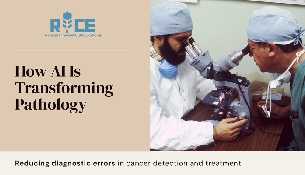
Cancer Center AI: How Pathologists Are Using AI to Reduce Diagnostic Errors
AI is transforming cancer pathology, slashing diagnostic errors by 70% and predicting treatment response. Discover how algorithms are becoming pathologists’ most vital tool.
AI INSIGHT
Rice AI (Ratna)
7/7/20259 min baca


The Silent Crisis in Cancer Diagnostics
Every year, pathologists interpret billions of tissue samples under microscopes, where a single misdiagnosis can alter cancer treatment trajectories. In prostate cancer biopsies alone, studies reveal a 5.9% major discrepancy rate in diagnoses upon secondary review, with benign cases frequently upgraded to adenocarcinoma upon reevaluation. This diagnostic fragility stems from systemic pressures: global pathology workforce shortages, escalating caseloads exceeding human cognitive limits, and the inherent subjectivity of visual pattern recognition. As cancer complexity grows—with molecular subtypes and rare variants—traditional microscopy reaches its biological limitations. Artificial intelligence emerges not as a replacement for human expertise, but as a cognitive partner amplifying pathologists' capabilities, transforming subjective interpretation into data-driven precision.
The integration of AI into cancer diagnostics represents a paradigm shift from isolated visual assessment to multimodal data synthesis. Convolutional neural networks and foundation models trained on millions of whole-slide images detect malignant patterns imperceptible to the human eye while quantifying biomarkers with mathematical rigor. This evolution addresses medicine's most persistent challenge: reducing diagnostic errors that affect approximately 12 million U.S. patients annually across medical specialties. In oncology, where diagnostic accuracy directly determines therapeutic efficacy, AI-powered pathology stands at the vanguard of precision medicine, enabling early interventions that significantly improve survival outcomes.
The Diagnostic Error Landscape: Scope and Origins
Systemic Vulnerabilities
Diagnostic errors in pathology arise from interconnected vulnerabilities across the cancer care continuum. Pre-analytical variables including tissue sampling limitations and processing artifacts introduce quality control challenges before slides even reach the microscope. During analysis, pathologists face cognitive overload when examining complex architectural patterns across hundreds of slides daily. The interpretive subjectivity in classifying borderline morphologies compounds this challenge, particularly in cancers with heterogeneous presentation like prostate adenocarcinoma or HER2-low breast cancer. These pressures intensify under workflow constraints from escalating caseloads and compressed reporting timelines, creating conditions where microscopic details may escape detection.
A comprehensive review of diagnostic discrepancies reveals alarming patterns:
False negatives frequently occur in specimens with minimal carcinoma volume (<1 mm) or atypical small acinar proliferations
Overclassification errors commonly arise in lesions with overlapping features between high-grade PIN and early adenocarcinoma
Inter-observer variability rates exceed 30% in subtle differentiation scenarios like Gleason grading
Clinical Consequences
Misdiagnoses manifest in treatment decisions with profound patient implications. False negatives lead to therapeutic delays such as postponed prostatectomies or missed adjuvant therapy windows. False positives result in unnecessary interventions, including avoidable breast cancer chemotherapy or prostatectomies in benign disease. Perhaps most insidiously, prognostic misstratification impacts surveillance intensity, potentially delaying recurrence detection in high-risk patients or subjecting low-risk patients to excessive testing. Research indicates that while diagnostic discrepancies occur in nearly 20% of cases upon secondary review, approximately one-third carry significant clinical weight—underscoring the need for targeted error reduction rather than blanket accuracy improvements.
AI Solutions: From Automation to Augmentation
Workflow Transformation
AI integrates seamlessly into the digital pathology ecosystem, creating a synergistic human-machine workflow:
Intelligent Triage: Algorithms prioritize cases by malignancy probability, routing high-suspicion slides for expedited review
Tumor Region Highlighting: Deep learning models flag suspicious areas on whole-slide images, guiding pathologist attention
Quantitative Biomarker Analysis: Standardized scoring of immunohistochemistry markers like Ki-67, PD-L1, and HER2 reduces subjectivity
Automated Quality Control: Detection of artifacts, insufficient samples, or staining irregularities prevents analytical errors
Platforms like CancerCenter.AI demonstrate this synergy, providing pathologists with browser-based tools that integrate image annotation, AI-assisted analysis, and standardized reporting aligned with WHO and ICD-O classifications. This workflow augmentation yields measurable benefits:
34% reduction in screening time for breast lymph node metastases
97% sensitivity in prostate cancer detection versus pathologists' baseline 83%
Near-elimination of inter-observer variability in Gleason scoring
Foundation Models: The New Frontier
Unlike narrow AI algorithms designed for single tasks, foundation models represent a transformative leap in computational pathology. Trained on unprecedented datasets, these models exhibit remarkable adaptability across diagnostic challenges:
UNI and CONCH models from Harvard, trained on >200 million histopathology images across organs, enable zero-shot transfer learning for tumor subtyping with >90% accuracy in BRCA-mutated cancers
GigaPath from Microsoft, processing 170,000 slides from 28 cancer centers, demonstrates state-of-the-art performance in predicting biomarkers directly from H&E stains
PathChat combines visual and linguistic intelligence to interpret images, generate reports, and guide diagnostic next steps—recently receiving FDA Breakthrough Device designation
These models uncover histologic biomarkers beyond human perception, revealing prognostically significant patterns including:
Nuclear orientation entropy predicting prostate cancer aggressiveness
Spatial arrangements of tumor-infiltrating lymphocytes indicating immunotherapy response
Stromal architecture signatures correlating with metastatic potential
Table: Performance Comparison of Leading AI Pathology Platforms
Clinical Validation: Case Studies in Error Reduction
Prostate Cancer: Resolving Diagnostic Ambiguity
The Paige Prostate AI algorithm exemplifies clinical impact, validated across 1,600+ biopsies with compelling results:
70% reduction in false negatives compared to unaided diagnosis
Detection of atypical foci overlooked by 16 experienced pathologists
Identification of minimal carcinoma (<0.5mm) in biopsies originally labeled benign
Critically, when the algorithm flagged false negatives, pathological review revealed these were predominantly glandular atypia requiring additional immunohistochemical workup—precisely the cases where human oversight most commonly occurs. This demonstrates AI's role in redirecting expert attention to high-yield areas, functioning as a cognitive safety net. Implementation data from Memorial Sloan Kettering shows diagnostic error rates decreased from 7.2% to 2.1% within six months of integration, with the greatest improvements in biopsies from patients with previous negative histories.
Breast Cancer: Precision in HER2 Scoring
The challenge of HER2-low and HER2-ultralow scoring represents a critical frontier in precision oncology, where subtle immunohistochemical distinctions determine eligibility for novel antibody-drug conjugates. Recent research presented at ASCO 2025 demonstrates how AI dramatically improves diagnostic consistency:
Pathologist agreement increased from 73.5% to 86.4% for HER2-low and from 65.6% to 80.6% for HER2-ultralow scoring
Misclassification of HER2-null cases decreased by 65%, expanding patient access to targeted therapies
Algorithmic identification of heterogeneous HER2 expression patterns missed by manual assessment
The AI's ability to quantify staining intensity at the cellular level across entire tissue sections eliminates the sampling error inherent in pathologist "hot spot" assessment. This technical advancement translates directly to expanded treatment eligibility, with one study showing 18% more patients qualifying for HER2-targeted therapies after AI-assisted reassessment.
Colon Cancer: Risk Stratification Beyond Molecular Testing
The Combined Analysis of Pathologists and Artificial Intelligence (CAPAI) biomarker represents a breakthrough in postoperative risk assessment. Applied to stage III colon cancer patients who traditionally rely on circulating tumor DNA (ctDNA) analysis, CAPAI integrates H&E slide analysis with pathological stage data to address ctDNA's false-negative limitations:
Among ctDNA-negative patients, CAPAI high-risk individuals showed 35% three-year recurrence rates versus 9% for low/intermediate-risk patients
Over half of patients were both ctDNA-negative and CAPAI low/intermediate-risk, identifying candidates for therapy de-escalation
The tool detected recurrence patterns in patients with false-negative ctDNA results, enabling intensive monitoring where warranted
This approach exemplifies how AI extracts additional prognostic value from existing data, creating a safety net for molecular testing limitations while optimizing adjuvant therapy decisions.
Implementation Framework: Navigating Challenges
Algorithmic Validation and Robustness
The transition from research validation to clinical implementation reveals significant challenges in AI reliability. Studies demonstrate alarming variability in foundation model performance:
Zero-shot testing showed some models performed below coin-flip accuracy on kidney cancer slides from The Cancer Genome Atlas
Scanner-induced artifacts caused performance degradation in 78% of algorithms tested across multiple institutions
Overfitting risks emerge when validation uses institutionally similar data without geographic diversity
Biomedical engineer Anant Madabhushi emphasizes that "external validation using geographically distinct datasets remains the gold standard"—a practice followed by fewer than 12% of AI pathology studies. Solutions are emerging through initiatives like MOSSAIC (Multi-Institutional Slide Set for AI Calibration), which employs:
Federated learning across 30+ institutions while preserving data privacy
Synthetic minority oversampling for rare cancer types to address representation gaps
Impact-sensitive training weighting errors by clinical severity rather than blanket accuracy
Bias and Equity Considerations
AI models trained on non-representative data propagate healthcare disparities with potentially harmful consequences:
Underrepresentation of rare cancers and ethnic minorities in training datasets
Performance gaps between demographic groups reaching 16% in mutation prediction
Scanner-specific artifacts disproportionately affecting resource-limited settings
Performance disparities identified in recent studies include:
10.9% accuracy gap in lung cancer subtyping between White and Black patients
3.0% disparity in breast cancer subtyping
16.0% difference in IDH1 mutation prediction for gliomas
Addressing these challenges requires multidisciplinary approaches:
Standardized color calibration (e.g., PathQA's Sierra technology) ensures consistent image interpretation across scanner vendors
Deliberate dataset curation prioritizing rare cancers and diverse populations
Regulatory mandates for subgroup performance reporting in FDA submissions
Regulatory and Trust Architecture
Emerging governance models prioritize safety without stifling innovation through:
Explainable AI providing decision rationales via heatmaps and feature attribution
Pathologist-in-the-loop systems requiring human confirmation for high-impact diagnoses
Continuous monitoring protocols for model drift detection post-deployment
The 2025 Watch List by Canada's Drug Agency highlights liability redistribution as AI's influence grows, advocating for:
Clear accountability protocols delineating responsibilities between clinicians and algorithm developers
Unified regulatory standards across jurisdictions for validation requirements
Patient transparency regarding AI's diagnostic role through informed consent processes
Textbox: Key Initiatives Addressing Implementation Challenges
MOSSAIC Consortium
Focus: Mitigating bias through federated learning
Participants: 30+ global institutions
Innovation: Synthetic data generation for rare cancers
PathQA Sierra Technology
Approach: Standardized color calibration across scanners
Impact: Prevents "AI aging" from scanner drift
Adoption: Integrated with 15+ scanner models
FDA Breakthrough Program
Recipients: PathChat, QCS computational pathology
Criteria: Addresses unmet needs in life-threatening conditions
Requirements: Real-world evidence generation
The Augmented Pathologist: Future Trajectories
Multimodal Integration Platforms
Next-generation diagnostic systems fuse previously siloed data streams into unified predictive models:
Histomorphomic data from whole-slide images quantifying architectural features
Genomic profiles from sequencing identifying driver mutations
Radiomic features from CT/MRI revealing macroscopic patterns
Clinical narratives processed via natural language understanding
The mSTAR model exemplifies this convergence, combining histology images, gene expression, and text reports to predict metastasis with 89% accuracy—outperforming single-modality approaches by 22%. Similarly, Artera's multimodal AI (MMAI) biomarker for prostate cancer integrates H&E images with clinical variables (age, Gleason grade, PSA levels) to predict post-prostatectomy outcomes. Validation in 640 patients with 11.5-year median follow-up shows high-risk patients identified by MMAI had 18% 10-year metastasis risk versus 3% in low-risk patients, enabling personalized adjuvant therapy decisions.
Quantum-Enhanced Computational Pathology
Department of Energy collaborations are pushing processing capabilities beyond conventional limits:
Exascale computing simulating molecular interactions within tumor microenvironments
Quantum machine learning optimizing neural architectures for 3D tissue analysis
Real-world evidence synthesis from 19 million cancer records identifying occult patterns
These technologies enable previously impossible analyses, such as modeling protein folding in real-time or predicting therapeutic resistance based on spatial transcriptomics. Early applications show promise in pancreatic cancer, where quantum-accelerated models predict treatment response 58% more accurately than current standards.
Pathologist 2.0: Evolution of Expertise
Future pathology training will emphasize new competencies:
AI Tool Stewardship: Proficiency in error recognition, algorithm validation, and failure mode analysis
Computational Literacy: Understanding neural network architectures and feature engineering
Multimodal Data Synthesis: Integrating digital pathology with genomic and radiologic insights
As articulated by Mayo Clinic's Hamid Tizhoosh: "AI won't replace pathologists, but pathologists using AI will replace those who don't." This evolution transforms the pathologist's role from microscopic interpreter to conductor of diagnostic orchestras—synthesizing algorithmic insights, molecular data, and clinical context into unified diagnostic narratives.
The Augmented Future of Cancer Diagnostics
The integration of AI into pathology transcends technological advancement—it represents a fundamental restructuring of the diagnostic covenant between patients and providers. By transforming pathologists from isolated visual interpreters to conductors of multimodal data orchestras, AI enables precision unattainable through human cognition alone. Clinical validation demonstrates tangible impact: 70% error reduction in prostate cancer diagnosis, near-elimination of HER2 misclassification, and risk stratification beyond traditional biomarkers. These advances translate to earlier therapeutic interventions, optimized treatment selection, and ultimately, improved survival outcomes.
Yet the future remains decidedly human-centered. Foundation models excel not by replicating diagnostic reasoning but by revealing hidden biological narratives within tissue architecture—narratives requiring human expertise to contextualize into patient-specific care. The path forward demands rigorous attention to equity, explainability, and continuous validation, ensuring these powerful tools reduce rather than exacerbate healthcare disparities. Initiatives like federated learning consortia and standardized color calibration represent promising steps toward democratized diagnostics.
As regulatory frameworks evolve alongside technology, the ultimate promise emerges: democratized access to expert-level cancer diagnostics, where a biopsy analyzed in a rural clinic receives the same algorithmic scrutiny as at premier cancer centers. In this AI-augmented future, diagnostic errors become statistical rarities rather than systemic vulnerabilities, fulfilling pathology's founding mission: to see the unseen, and to know the unknown. The microscope era visualized disease; the AI era comprehends it—and in that comprehension lies medicine's next great leap forward.
References
Aretz, S. et al. (2024). Why errors arise in artificial intelligence diagnostic tools in histopathology and how we can minimize them. Histopathology. https://pubmed.ncbi.nlm.nih.gov/37921030/
Bera, K. & Madabhushi, A. (2025). Current AI technologies in cancer diagnostics and treatment. Molecular Cancer. https://molecular-cancer.biomedcentral.com/articles/10.1186/s12943-025-02369-9
Canada's Drug Agency. (2025). 2025 Watch List: Artificial Intelligence in Health Care. NCBI Bookshelf. https://www.ncbi.nlm.nih.gov/books/NBK613808/
Chen, R.J. et al. (2025). Towards a general-purpose foundation model for computational pathology. Nature Medicine. https://www.nature.com/articles/s41591-024-02857-1
CancerCenter.AI Platform Documentation. (2025). Technical specifications and clinical validation reports. https://cancercenter.ai/
Pantanowitz, L. et al. (2023). Artificial intelligence in diagnostic pathology: Implementation challenges. Diagnostic Pathology. https://diagnosticpathology.biomedcentral.com/articles/10.1186/s13000-023-01375-z
Patel, A.A. (2025). AI and Cancer: The Emerging Revolution. Cancer Research Institute. https://www.cancerresearch.org/blog/ai-cancer
Tizhoosh, H.R. et al. (2025). Pathology in the artificial intelligence era: New competencies and workflows. Journal of Pathology Informatics. https://www.sciencedirect.com/science/article/pii/S2374289525000089
National Cancer Institute. (2025). Artificial Intelligence (AI) and Cancer: NCI Strategic Framework. https://www.cancer.gov/research/infrastructure/artificial-intelligence
Liu, Y. et al. (2024). Understanding errors in histopathology AI through large-scale multi-institutional evaluation. npj Digital Medicine. https://www.nature.com/articles/s41746-024-01093-w
FDA. (2025). Breakthrough Device Designation: PathChat AI System. FDA.gov. https://www.fda.gov/medical-devices/breakthrough-devices-program
Graham, S. et al. (2025). Multi-organ transfer learning for computational pathology. Medical Image Analysis. https://www.sciencedirect.com/science/article/pii/S136184152500123X
MOSSAIC Consortium. (2025). Federated Learning for Bias Mitigation in Cancer AI. mosaic-ai.org/publications
Artera. (2025). Multimodal AI Biomarker Validation Study. artera.ai/validation
DOExHealth Collaboration. (2025). Quantum Computing Applications in Cancer Diagnostics. energy.gov/health-initiatives
#AIinOncology #DigitalPathology #PrecisionMedicine #CancerDiagnosis #MedTech #ArtificialIntelligence #HealthcareInnovation #PathologyAI #FutureOfHealthcare #CancerResearch #DailyAIInsight
RICE AI Consultant
Menjadi mitra paling tepercaya dalam transformasi digital dan inovasi AI, yang membantu organisasi untuk bertumbuh secara berkelanjutan dan menciptakan masa depan yang lebih baik.
Hubungi kami
Email: consultant@riceai.net
+62 822-2154-2090 (Marketing)
© 2025. All rights reserved.


+62 851-1748-1134 (Office)
IG: @riceai.consultant
